




Vascular Ultrasound • RPVI Preparation • 2026
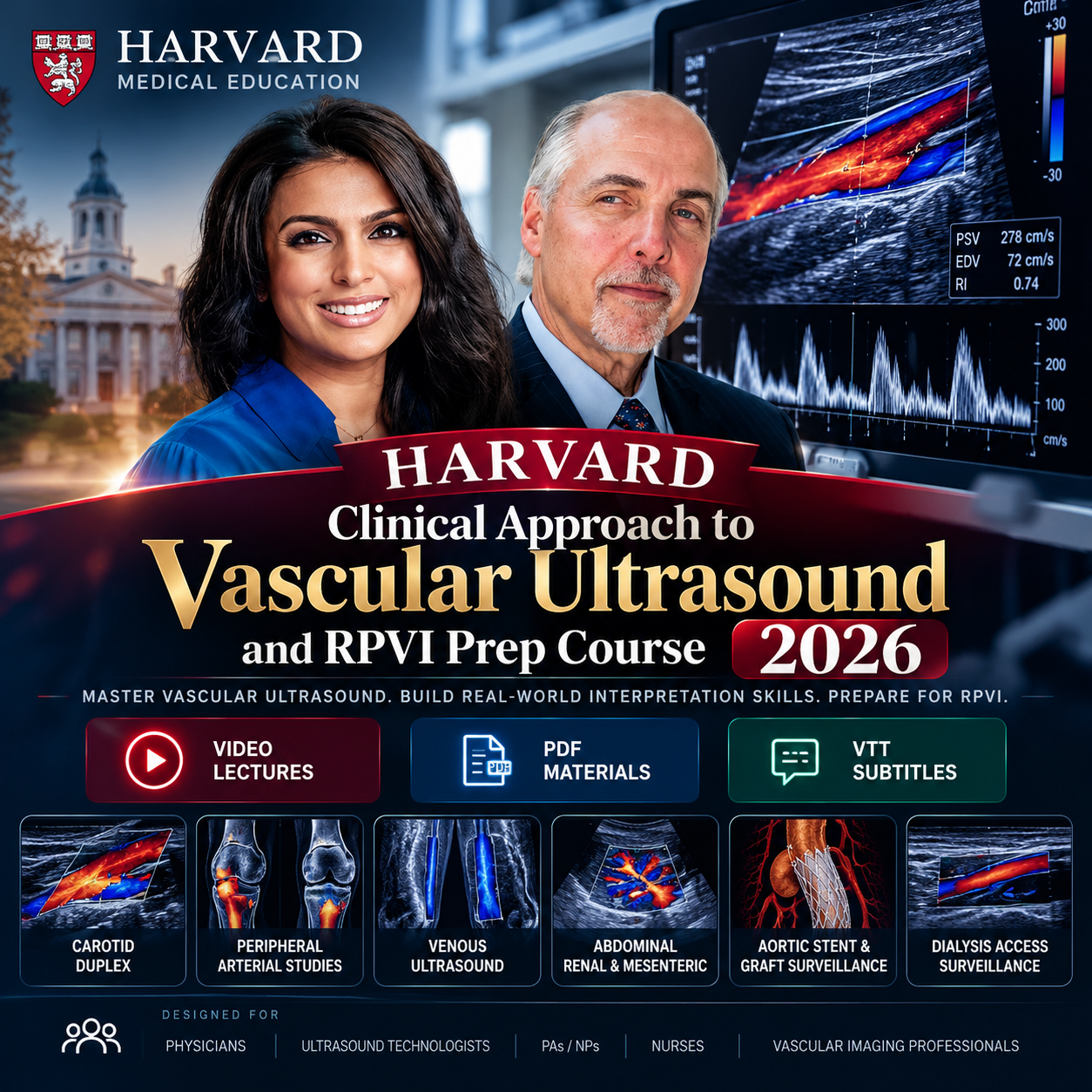
Harvard Clinical Approach to
Vascular Ultrasound
and RPVI Prep Course 2026
Build practical confidence in vascular ultrasound performance, Doppler interpretation, velocity criteria, vascular pathology, case analysis, and RPVI-focused review.
Videos
Recorded Lectures
PDFs
Course Materials
VTT
Subtitle Files
June 5–6
2026 Course Dates
Peripheral Arterial
Carotid Duplex
Venous Ultrasound
Abdominal Vascular
Graft Surveillance
Dialysis Access
RPVI Prep
Carotid Duplex
Venous Ultrasound
Abdominal Vascular
Graft Surveillance
Dialysis Access
RPVI Prep
Course Overview
From Ultrasound Physics to Confident Vascular Interpretation
Harvard Clinical Approach to Vascular Ultrasound and RPVI Prep Course 2026 is designed to strengthen both the performance and interpretation of vascular ultrasound across peripheral arterial, venous, cerebrovascular, abdominal, graft, dialysis access, and post-intervention studies.
The course combines ultrasound physics, vascular imaging technique, Doppler interpretation, velocity criteria, vascular pathology, case-based learning, RPVI-focused review, and vascular laboratory development to connect imaging findings with real clinical decisions.
Clinical Learning Framework
Acquire → Interpret → Apply → Decide
01
Acquire
Understand ultrasound physics, machine controls, scanning principles, and image acquisition.
02
Interpret
Recognize Doppler patterns, vascular pathology, abnormal flow, and clinically relevant findings.
03
Apply Criteria
Use velocity and diagnostic criteria in stenosis, grafts, dialysis access, and endovascular surveillance.
04
Decide
Connect vascular ultrasound findings to patient management and real-world vascular laboratory practice.
Major Clinical Domains
A Broad Review of Vascular Ultrasound Practice
Peripheral Arterial Imaging
Upper and lower extremity studies, peripheral artery disease, tibio-pedal access, and bypass graft assessment.
Carotid & Cerebrovascular Imaging
Carotid duplex, vertebral and subclavian ultrasound, transcranial Doppler, and cerebrovascular interpretation.
Venous Ultrasound
Venous insufficiency, thrombosis, obstruction, pelvic congestion syndrome, procedures, and case interpretation.
Abdominal Vascular Ultrasound
Renal and mesenteric artery disease, abdominal ultrasound, and vascular diagnostic applications.
Graft & Access Surveillance
Bypass grafts, dialysis access, aortic stent graft surveillance, and deep venous arterialization grafts.
RPVI-Focused Review
High-yield pathology, ultrasound physics, interpretation principles, velocity criteria, and commonly tested concepts.
Why This Review Matters
The Waveform Is Only Part of the Story
Interpretation
Move beyond image acquisition to understand findings, pathology, velocity changes, and clinical meaning.
Case-Based Learning
Review practical vascular cases designed to connect ultrasound findings with real patient situations.
RPVI Preparation
Reinforce important physics, pathology, interpretation, and high-yield concepts relevant to RPVI preparation.
Learning Objectives
Strengthen Both Technique and Interpretation
Demonstrate clinical interpretation of arterial and venous noninvasive studies in peripheral, cerebrovascular, and abdominal vascular systems.
Apply velocity criteria for grading stenosis in vascular territories, dialysis grafts, endovascular interventions, and bypass grafts.
Review essential ultrasound physics and technology as they apply to vascular diagnostics.
Reinforce high-yield concepts commonly encountered in Registered Physician in Vascular Interpretation preparation.
Understand core considerations involved in developing and implementing a vascular laboratory program.
Complete Curriculum
29 Sessions Across Vascular Ultrasound
00 — Introduction
01 — Basics of Physics in Vascular Imaging
02 — Understanding the Ultrasound Machine
03 — Peripheral Arterial Studies – Upper
04 — Peripheral Arterial Studies – Lower
05 — Abdominal Ultrasound
06 — Panel Discussion
07 — Renal and Mesenteric Artery Disease
08 — Carotid Duplex Ultrasonography
09 — Subclavian and Vertebral Ultrasonography
10 — Cerebrovascular Intracranial (Transcranial Doppler)
11 — High-Yield RPVI Pathologies
12 — Panel Discussion
13 — Bypass Graft Surveillance
14 — Aortic Endovascular Stent Graft Surveillance
15 — Dialysis Access Surveillance and Case Presentations
16 — Tibio-Pedal Access
17 — Panel Discussion
18 — Venous Ultrasound I – Venous Insufficiency & Office Procedures
19 — Venous Ultrasound II – Thrombosis, Obstruction & Pelvic Congestion
20 — Venous Case Presentation
21 — Wound Healing in Vascular Surgery
22 — Carotid Arterial Case Presentations
23 — DEEPER OUS & DEEPER REVEAL Trial Updates
24 — Peripheral Artery & Bypass Graft Case Presentations
25 — Surveillance of Deep Venous Arterialization Grafts
26 — Deep Venous Arterialization
27 — Pedal Acceleration Time (PAT): Technique, Criteria, Interpretation & Clinical Application
28 — Panel Discussion
Who Should Participate?
Designed for Clinicians and Vascular Imaging Professionals
Primary Care Physicians
Specialty Physicians
Ultrasound Technologists
Physician Assistants
Nurse Practitioners
Nurses & Allied Professionals
Course Package
Included Educational Materials
VIDEO
Recorded Lectures
PDF
Course Materials
VTT
Subtitle Files
Vascular Ultrasound • Doppler • Velocity Criteria • RPVI Review
See the Waveform. Apply the Criteria. Strengthen the Interpretation.
Review the major vascular ultrasound studies, interpretation principles, clinical applications, surveillance strategies, and RPVI-focused concepts in one structured course.
Certificate Notice:
This purchase includes educational course materials only. Our store does not issue CME/CE credit, an RPVI credential, official accreditation, or a certificate of completion.
This purchase includes educational course materials only. Our store does not issue CME/CE credit, an RPVI credential, official accreditation, or a certificate of completion.


Reviews
There are no reviews yet.